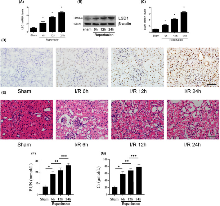
FIGURE 1.

LSD1 expression was elevated in mice after renal IRI. All mice were subjected to ischaemia 30 min and reperfusion 6, 12 and 24 h, respectively. (A) LSD1 mRNA level was elevated after renal IRI. (B‐C) LSD1 protein level was elevated after renal IRI, and the quantification also was shown. (D) The immunohistochemical staining of LSD1 was examined in renal tissues at 6, 12 or 24 h of reperfusion time (×400; scale bars = 40 μm). (E) The morphological changes of the renal tissues detected by H&E staining at 6, 12 or 24 h of reperfusion (×400; scale bars = 40 μm)(F–G)The levels of Cr and BUN were detected after renal IRI (n = 8) The results were expressed as mean ± standard error of mean (SEM). *p < 0.05, when compared with the sham group. **p < 0.05, when compared with the IR 6 h group. ***p < 0.05, when compared with the IR 12 h group
