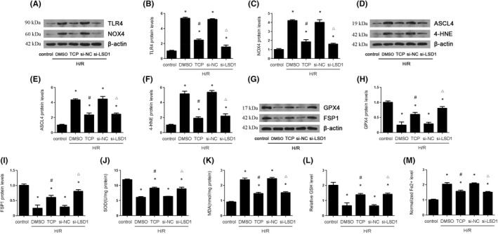
FIGURE 5.

LSD1 inhibition decreased TLR4/NOX4, ferroptosis and oxidative stress that were caused by H/R in HK‐2 cells. The HK‐2 cells were subjected to hypoxia 12 h and reoxygenation 6 h. The cells were pretreated with TCP or transfected with si‐NC or si‐LSD1 for 24 h and then subjected to H/R. (A–C) The expression of TLR/NOX4 pathway was examined by Western blot, and the quantification was also shown. (D–I) The regulatory effect of LSD1 on ASCL4, 4‐HNE, GPX4 and FSP1 expression in HK‐2 cells after H/R, and quantification. (J) The regulation of LSD1 on SOD activity in HK‐2 cells after H/R. (K) The regulation of LSD1 on MDA content in HK‐2 cells after H/R. (L) The regulation of LSD1 on GSH level in HK‐2 cells after H/R. (M) The regulation of LSD1 on Fe2+ level in HK‐2 cells after H/R (n = 8). The results were expressed as mean ± SEM. *p < 0.05, when compared with the control group. #p < 0.05, when compared with the H/R + DMSO group. △p < 0.05, when compared with the H/R + si‐NC group
