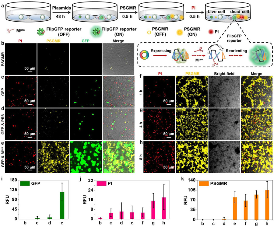
Figure 3. Validation of PSGMR via plasmids and reporter.
(a) The experimental scheme of different plasmids transfected HEK 293T cells after incubation with PI, and PSGMR. The enlarged portion shows the imaging mechanism of FlipGFP. CLSM images of the HEK 293T cells with (b) PSGMR, (c) Mpro-related FlipGFP reporter plasmid and PSGMR, (d) Mpro-related FlipGFP reporter plasmid, PR8 plasmid and PSGMR, (e) Mpro-related FlipGFP reporter plasmid, Mpro plasmid and PSGMR, and Mpro plasmid and PSGMR for (f) 1h, (g) 4h, and (h) 8h. Average fluorescence intensities of (i) GFP, (j) PI and (k) PSGMR in each panel. The FlipGFP and PSGMR are activated in the green and yellow channel when cells are transfected with the Mpro plasmid. The PI is activated in the red channel when cells are dead.

