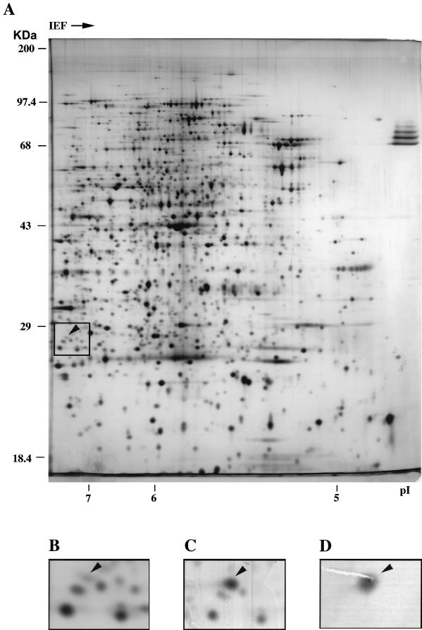
FIG. 3.
Identification of UMP kinase in the E. coli proteome by two-dimensional gel electrophoresis. (A) Fifty micrograms of soluble protein from strain BL21(DE3) was silver stained after two-dimensional gel electrophoresis. The molecular mass markers are indicated on the left of the gel, and the isoelectric point scale is on the bottom. (B) Enlarged portion of panel A, in which the position of UMP kinase (arrowhead) and surrounding protein spots can be easily recognized. (C) A multicopy plasmid harboring the pyrH gene was introduced into strain BL21(DE3), and a 10-μg sample of soluble protein was analyzed as for panel A. An enlarged portion is shown, as in panel B. (D) One hundred micrograms of soluble protein from strain NM554 was separated by two-dimensional gel electrophoresis, and UMP kinase was immunodetected with polyclonal rabbit antiserum as described in Materials and Methods. An enlarged portion is shown, with the same magnification as in panels B and C.

