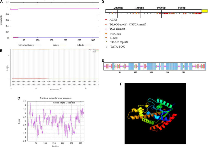
FIGURE 1.
The characterization of the ZmIPT2 protein. (A) Transmembrane domain prediction of ZmIPT2. (B) Prediction of signal peptide and splice siteS. (C) Hydrophobicity analysis of ZmIPT2 coding protein. (D) Analysis of flanking sequence of ZmIPT2 gene. (E) Secondary structure analysis of the ZmIPT2. The helix (h), extended strand (e), coil(c), and turn (t) are indicated in different color as blue, red, yellow, and green, respectively. (F) 3-D structure model of ZmIPT2.

