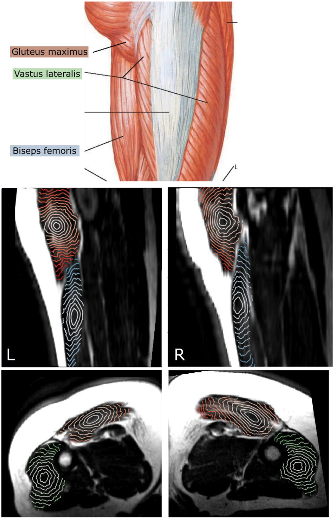
Figure 3.

Top: muscles of the thigh, gluteus maximus, biseps femoris, vastus lateralis. (Adapted from (27)). Middle: sagittal view of the T1-weighted MRI with isosurfaces of shortest distance calculated within the gluteus maximus (orange) and biseps femoris (blue) muscles starting from a point in the center. Bottom: axial view with the isosurfaces calculated within gluteus maximus (orange) and vastus lateralis (green) muscles.
