
Figure 1:
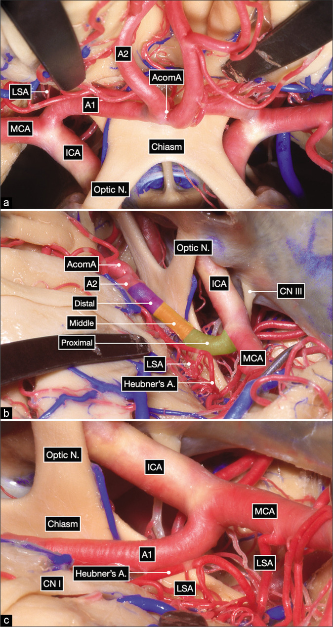
Anatomic dissection in a formalin-fixed and silicone-injected cadaveric head. (a) Anterior view after elevating the frontal lobes showing the bilateral optic nerves, optic chiasm and bilateral carotid bifurcations. The proximal, middle and distal subsegments of the first (precommunicating or A1) segment of the anterior cerebral artery (ACA) are delineated on the right side. (b and c) Surgeon’s perspective in a right pterional craniotomy after exposing the internal carotid artery bifurcation. The A1 subsegments are colored as follows: proximal in green, middle in yellow and distal in purple.