Figure 3:

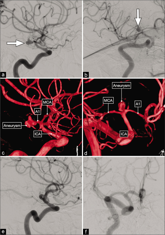
Lateral (a) and anterior oblique (b) views of a diagnostic cerebral angiogram showing a right-sided, dysmorphic, posteriorly-projecting A1 segment aneurysm (white arrow). (c and d) Three-dimensional reconstructions that better delineate the exact origin, neck and projection of this aneurysm on the proximal A1 subsegment. (e and f) Postoperative cerebral angiograms showing complete occlusion of the aneurysm and good flow across the ACA.
