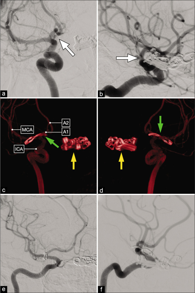
Figure 5:

Anteroposterior (a) and lateral (b) views of a diagnostic cerebral angiogram showing a right-sided, posteriorly-projecting A1 segment aneurysm (white arrow). Tandem clips occluding a left-sided paraclinoid ICA aneurysm are also noted. (c and d) Postoperative 3D reconstruction showing a curved clip occluding the right-sided A1 segment aneurysm (green arrow) and multiple tandem clips occluding the left-sided paraclinoid aneurysm (yellow arrow). (e and f) Postoperative diagnostic cerebral angiogram demonstrating complete aneurysm clip occlusion.
