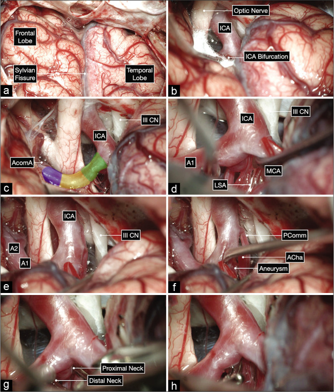
Figure 6:

Intraoperative microphotographs of the second case. (a) A right pterional craniotomy was performed to expose the right Sylvian fissure and adjacent frontal and temporal lobes. (b and c) A wide Sylvian fissure and arachnoid dissection exposed the optic nerve, ICA bifurcation, and all subsegments of the A1: proximal (in green), middle (yellow), and distal (purple). (d-h) Meticulous arachnoid dissection skeletonized the lenticulostriate arteries (LSA) and exposed the aneurysm projecting posteriorly and medially. The proximal and distal neck of the aneurysm were also exposed and a clip was applied parallel to the parent A1 using a trajectory superior to the MCA.
