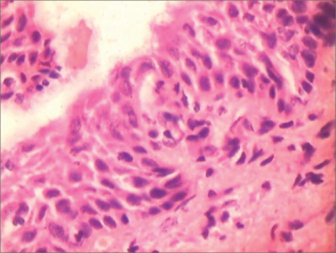
Figure 31:

Biopsy from a benign atrophic epithelium showing presence of hyperchromatic but generally uniform nuclei. The epithelium is thin with maintained polarity. The nuclear appearance is predominantly smudgy, hyperchromatic without recognizable nucleoli, and internal structure is no longer discernible. Colposcopic impression was HSIL.
