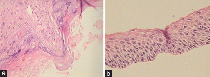
Figure 8:

(a and b) Spiky (a) condyloma. The surface of the spikes shows hyperkeratosis, parakeratosis, and koilocytes beneath (×10). (b) Flat condyloma lacks the papillary architecture of an exophytic lesion (×10). Both these generally present as subclinical condylomas and are detected only when they turn acetowhite after application of 5% acetic acid or during colposcopy.
