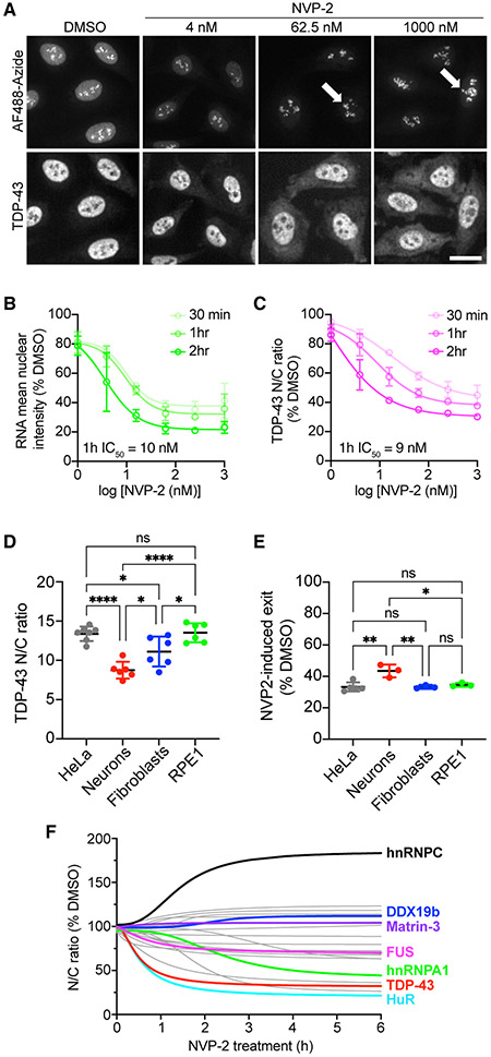
Figure 1. NVP2-induced RNA pol II inhibition promotes TDP-43 nuclear export.
(A) Nascent RNA labeled with AF488-picolyl azide via 5-EU/click chemistry (top) and TDP-43 IF (bottom) in HeLa cells treated with NVP2 for 1 h. Arrows indicate rRNA puncta unaffected by NVP2. Scale bar, 25 μm.
(B and C) Nascent RNA (AF488) mean nuclear intensity (B) and TDP-43 nuclear/cytoplasmic ratio (N/C) (C) expressed as percentage of DMSO control. Mean ± SD is shown for >2,000 cells/group in each of three biological replicates. IC50 at 1 h was calculated by non-linear regression. See also Figure S1.
(D) Steady-state TDP-43 N/C in HeLa cells, mouse primary cortical neurons, human HFF1 fibroblasts, and human retinal pigment epithelial (RPE1) cells fixed and immunostained with mouse monoclonal anti-TDP-43. Mean ± SD is shown for 6–7 biological replicates. See also Figure S2.
(E) NVP2-induced shift in TDP-43 N/C, shown as percentage of DMSO control, following 3 h with 250 nM NVP2. Mean ± SD is shown for 3–5 biological replicates.
(F) RBP N/C in HeLa cells treated with 250 nM NVP2 for 30 min to 6 h. Curves were fit by non-linear regression using the mean of 3–4 biological replicates per RBP. See also Figure S3.
In (D), (E), and (F), >2,000 cells/replicate for cell lines and 60–80 cells/replicate for neurons. ns, not significant; *p < 0.05, **p < 0.01, ****p < 0.0001 by one-way ANOVA with Tukey’s test.

