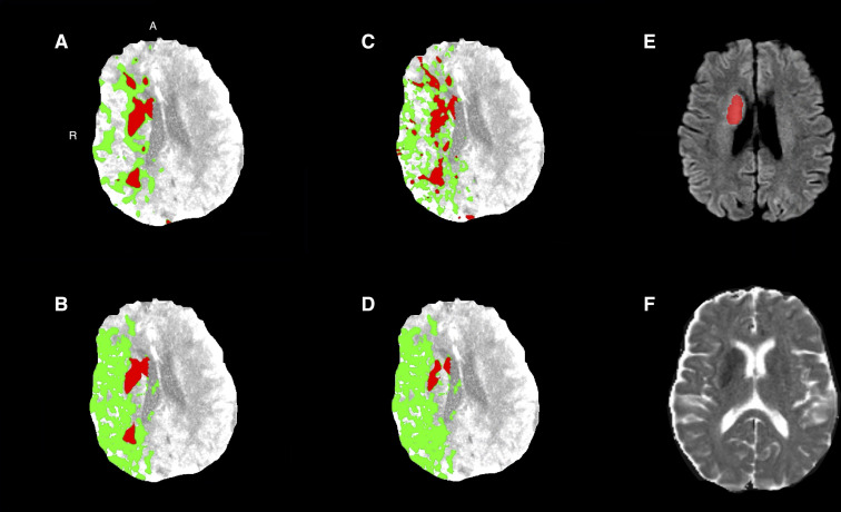
Fig 3. Baseline CTP of a patient with a right-sided M1 occlusion with successful reperfusion (eTICI 3).
The ischemic core (red) and penumbra (green) for (a) approach 1, (b) approach 2, (c) approach 3, and (d) approach 4. (e) Follow-up DWI acquired at 17 hours after baseline imaging and (f) follow-up DWI image with follow-up infarct segmentation (red). A = anterior; CTP = CT perfusion; DWI = diffusion weighted imaging; MRI = magnetic resonance imaging; R = right.

