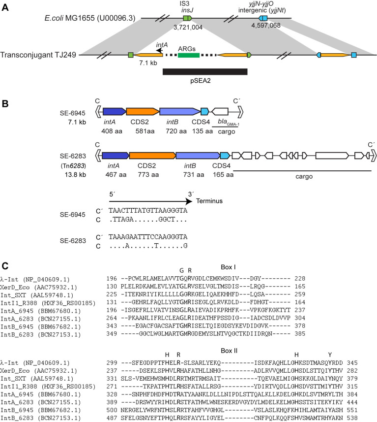
Fig 1. Genetic features of SEs.
(A) Schematic representation of the 7.1 kb element and pSEA2 insertion locations in E. coli TJ249. Yellow pentagons are the 7.1 kb element. The sequence from pSEA2 is indicated by a horizontal line. (B) Upper panel: Genetic organization of SE-6283 and SE-6945. Lower panel: Nucleotide sequences of imperfect inverted repeat motifs C and C’. Dots indicate that bases in C are identical to those in C’. (C) Presence of R-H-R-Y motif in IntA and IntB. IntA and IntB sequences are aligned with sequences of known tyrosine recombinases based on secondary structure prediction using PROMAL 3D [34]. The aligned regions corresponding to box I and box II in [35] are shown.

