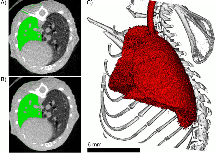
Figure 3.
Representative cross section of CT data sets of the chest region of a mouse reconstructed (A) without RG and (B) with RG and frame averaging. In green the segmented lung using the same threshold is partially overlaid. Clearly, in (B) both, less motion artifacts and a sharper delineation of the lung towards the rib cage are observed. This allows to study the anatomical shape of the lung in 3D as demonstrated in (C). Note, that the interface between lung and heart was not improved since no gating was performed for the movement of the heart. The figure was generated using imagej 1.53f www.imagej.nih.gov/ij/, gimp 2.10.28 www.gimp.organd scry (proprietary 3D render software).

