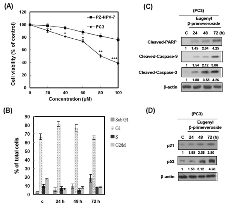
Fig. 4.
Eugenyl β-primeveroside induced cell cycle arrest and apoptosis in PC3 cells. (A) PC3 and PZ-HPV-7 cells were treated with various concentrations of eugenyl β-primeveroside at 37°C for 48 hours. The effect on cell growth was examined by 3-(4,5-dimethylthiazol-2-yl)-2,5-diphenyl tetrazolium bromide assay, and the percentage of cell proliferation was calculated by defining the absorption of cells without eugenyl β-primeveroside as 100%. This experiment was repeated three times. Bars represent the standard deviation. Asterisks indicate that the value is significantly different from that of the control (*p < 0.05; **p < 0.01; ***p < 0.001). (B) PC3 cells were treated with 80μM of eugenyl ββ-primeveroside for the indicated duration and analyzed for PI-stained DNA content by flow cytometry. The indicated percentages are the mean of three independent experiments, each in duplicate. Bars represent the standard deviation. (C) PC3 cells were treated with vehicle (dimethyl sulfoxide), eugenyl β-primeveroside (80μM) for the indicated time. Cells were then harvested and lysed for the detection of cleaved PARP, Caspase-9, Caspase-3 and β-actin protein expression. (D) PC3 cells were treated with vehicle (dimethyl sulfoxide), eugenyl β-primeveroside (80μM) for the indicated time. Cells were then harvested and lysed for the detection of p21, p53 and β-actin protein expression. Western blot data presented are representative of those obtained in at least three separate experiments. Immunoblots were quantified, and relative expression to control is indicated.

