FIG. 4.
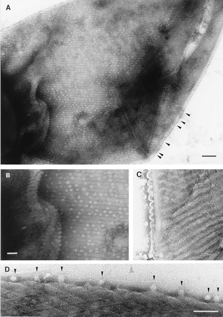
Transmission electron micrographs of Oscillatoria sp. strain FT1. Actively motile samples were crushed between glass slides and negatively stained. (A) The cell walls are covered with regularly arranged circular structures approximately 20 nm in diameter, which are unstained except at the center. These structures are located between the electron-dense lines that separate the fibrils and appear not to be pores, because they can be lost from the surface and seen free, external to the cell (arrowheads here and in panel D). Bar, 200 nm. (B) Shown is an enlarged view of part of panel A, revealing the circular structures that follow the line of each fibril. Bar, 100 nm. (C) The fibrils are separated by an electron-dense line bounded on each side by electron-transparent regions. The former is caused by the accumulation of stain in the cleft in the outer membrane, where it dips between each row of fibrils, and the latter is the unstained membrane itself, on either side of the cleft. These corrugations in the outer membrane can be seen to the left of the micrograph (and are illustrated in Fig. 7). Bar, 100 nm. (D) Enlarged view of part of the edge of the cell from the top left corner of panel A, showing the unstained, 20-nm-diameter circular structures (arrowheads) external to the cell. Bar, 100 nm.

