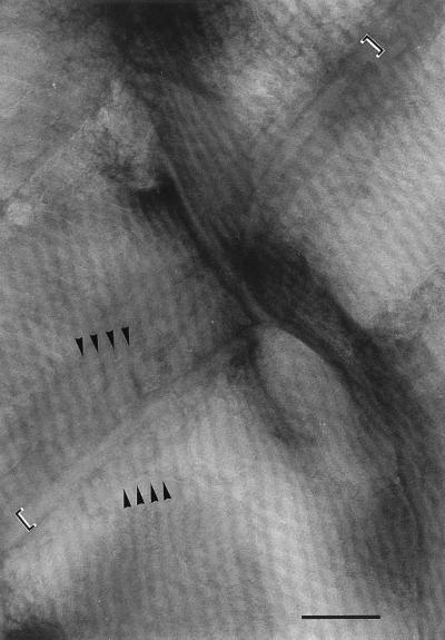
FIG. 5.
Enlarged view of the top left corner of Fig. 2. The cell septum runs from the bottom left to the top right of the photograph. Each fibril is seen as a pale line bounded by two parallel dark lines, and at the point at which they cross the cell septum they appear to pass beneath a narrow band (indicated by the square brackets) that follows the line of the septum. The continuation of three of the fibrils, on either side of the septum, is shown by the arrowheads indicating the dark lines between the fibrils. Bar, 200 nm.

