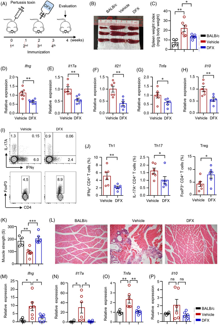
FIGURE 6.

Iron chelation suppressed CD4+ T‐cell response during autoimmune myositis. (A) Scheme of the mouse experiment. Experimental autoimmune myositis (EAM) was induced as described in Section 4. Mice were first injected with 500‐ng pertussis toxin intraperitoneally and then immunized with myosin (1 mg) emulsified with complete Freund's adjuvant four times at 1‐week interval. The EAM mice were fed with deferasirox (DFX) (30 mg/kg/d) dissolved in the drinking water after the second immunization for 2 weeks. (B and C) Spleen weight index as calculated spleen weight (mg) to body weight (g) 4 weeks after the primary immunization. (D)–(H) Ifng, Il17a, IL21, Tnfa and Il10 gene expression in the draining lymph nodes were measured by quantitative real‐time PCR (qPCR). (I and J) Interferon gamma (IFNγ)‐, interleukin (IL)‐17A‐ and FoxP3‐producing CD4+ T‐cells in draining lymph nodes were measured by flow cytometry. Representative counter plots were shown. (K) Muscle strength of the EAM mice treated with DFX or vehicle. (L) Muscle (quadriceps) sections of mice treated with DFX or vehicle were stained with hematoxylin and eosin. Representative images are shown. Original magnification: 100×. (M)–(P) RNA was extracted from muscle tissue of quadriceps. Transcripts of Ifng, Il17a, Tnfa and Il10 in the muscle tissues were quantified by qPCR. All data are mean ± SEM. n = 6. *p < .05, **p < .01, by one‐way ANOVA followed by adjustments for multiple comparisons in panels (C), (K), (M)–(P) and Student's t‐test in panels (D)–(H) and (J). ns, not significant.
