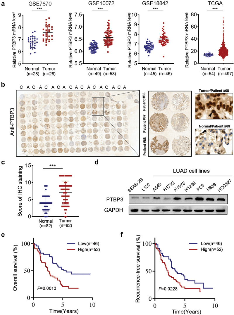
Figure 1.

PTBP3 is upregulated in LUAD patients and indicates poor prognosis. (a) PTBP3 mRNA expression in normal lung tissues and primary LUAD tissues revealed by four different public databases, including GSE7670, GSE10072, GSE18842 and TCGA. ***P < 0.001 by Student’s t-test. (b) IHC staining showed that expression of PTBP3 in adjacent nontumor tissues and LUAD tissues (magnification ×400). (c) IHC scores of PTBP3 expression in adjacent nontumor tissues and primary LUAD tissues (n = 82). ***P < 0.001 by Student’s t-test. (d) Western-blot analysis of PTBP3 expression in human alveolar and bronchial epithelial cell lines (L132 and BEAS-2B), LUAD cell lines (A549, H1299, H1975, H1792, PC9, H838 and HCC827). GAPDH was used as an internal control. (e and f) Ninety-eight LUAD patients were divided into low (n = 46) and high (n = 52) PTBP3 expression groups as described in Materials and Methods. Kaplan-Meier analysis of overall survival (e) and recurrence-free survival (f) in LUAD patients stratified by PTBP3 protein level. Log-rank Test was used to analyze differences between groups.
