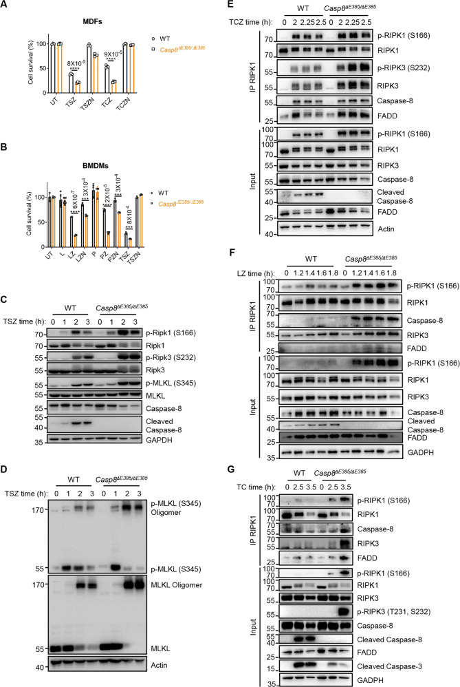
Fig. 3. The CASP8(ΔE385) promotes necroptosis in vitro.
A Primary WT and Casp8ΔE385/ΔE385 MDFs were treated with TNF-α (20 ng/ml)+Smac (1 μM)+zVAD (20 μM) (TSZ) and TNF-α + Smac + zVAD + Nec-1 (30 μM) (TSZN) for 6.45 h, TNF-α + CHX (20 μg/ml)+zVAD (20 μM) (TCZ) and TNF-α + CHX + zVAD + Nec-1 (30 μM) (TCZN) for 4.45 h. Bars, mean ± SD. P values above the asterisk (unpaired, two-tailed t test) ****p < 0.0001. B Primary WT and Casp8ΔE385/ΔE385 bone marrow derived macrophages (BMDMs) were treated with LPS (100 ng/ml), LPS + zVAD (20 μM) (LZ), LPS + zVAD + Nec-1 (30 μM) (LZN), poly(I:C) (100 μg/ml), poly(I:C) + zVAD (20 μM) (PZ), poly(I:C) + zVAD + Nec-1 (30 μM) (PZN), TNF-α + Smac + zVAD (TSZ), TNF-α + Smac + zVAD + Nec-1 (TSZN) for 3 h. Bars, mean ± SD. P values above the asterisk (unpaired, two-tailed t test) ***p < 0.001, ****p < 0.0001. C Immunoblotting of the indicated protein expression in primary WT and Casp8ΔE385/ΔE385 MDFs which were treated with TNF-α (20 ng/ml) +Smac (1 μM) +zVAD (20 μM) (TSZ) for the indicated time. D Immunoblotting of primary WT and Casp8ΔE385/ΔE385 MDFs which were treated with TNF-α (20 ng/ml) +Smac (1 μM) +zVAD (20 μM) (TSZ) for the indicated time. E WT and Casp8ΔE385/ΔE385 MDFs were treated with TNF-α (40 ng/ml)+CHX (40 μg/ml)+zVAD (50 μM) for the indicated time, complex II was immunoprecipitated using anti-RIPK1, the recruitment of RIPK3, FADD and caspase-8 were detected by western blotting. F Primary WT and Casp8ΔE385/ΔE385 BMDMs were treated with LPS (200 ng/ml)+zVAD (40 μM) followed by western blot and immunoprecipitation. G Primary WT and Casp8ΔE385/ΔE385 MDFs were treated with TNF-α (40 ng/ml)+CHX (40 μg/ml) followed by western blot and immunoprecipitation.

