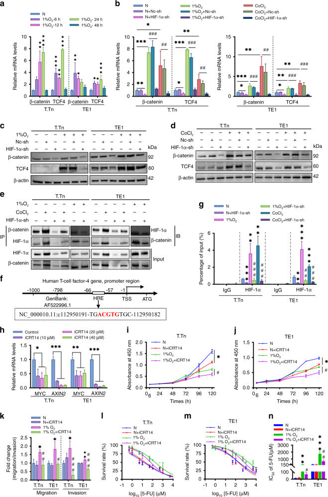
Fig. 5. Crosstalk between HIF-1α and Wnt/β-catenin signalling pathway in ESCC cell lines.
a β-catenin and TCF4/TCF7L2 mRNA levels in T.Tn and TE1 cells at different time points under 1% O2 exposure. b The mRNA levels of β-catenin and TCF4/TCF7L2 were detected T.Tn and TE1 cell lines with or without HIF-1α shRNA transfection in the presence of 1% O2 (T.Tn, 24 h; TE1, 12 h) or CoCl2 (100 μM, 24 h). Data were normalised to normoxia. c, d Western blot assays for β-catenin and TCF4/TCF7L2 protein expression were performed in the indicated cells as described above. e The physical association between HIF-1α and β -catenin in indicated T.Tn and TE1 cells is based on co-immunoprecipitation (Co-IP) blotting. f Schematic representation of the human TCF4/TCF7L2 promoter region. g The relative abundance of HIF-1α binding to the TCF4/TCF7L2 promoter in indicated T.Tn and TE1 cells was measured by ChIP. ChIP-qRT-PCR quantitation was shown as % relative to input, IgG was used as a negative control. h The relative mRNA levels of MYC and AXIN2 in iCRT14-treated T.Tn and TE1 cells at the indicated concentrations under normoxic conditions. Effects of iCRT14 (10 μM, 24 h pretreatment) on cell proliferation (i, j), migration/invasion (k), 5-fluorouracil (5-FU) resistance (l, m) in T.Tn and TE1 cells cultured in normoxic and hypoxic (1% O2) environments. n Corresponding IC50. All data represent mean ± SD of three independent experiments. *P < 0.05, **P < 0.01, ***P < 0.001 vs normoxic baseline. #P < 0.05, ##P < 0.01, ###P < 0.001 vs hypoxic (1% O2 or CoCl2) baseline. TSS transcription starting site, HRE hypoxia-responsive element, N normoxia, Nc-sh negative control shRNA, HIF-1α-sh HIF-1α-shRNA.

