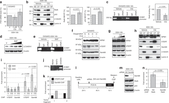
Fig. 4. hTERT and Sam68 contribute to NFκB binding to ICAM1 under SSH.
a Real-time RT-PCR analysis of FVII mRNA expression in cells exposed to CQ under the indicated conditions. b Western blot analysis of RelA and p50 expression in cytoplasmic and nuclear fractions of OVSAYO cells. NE, CP, and NP are the nuclear extract, cytoplasmic extract, and nuclear pellet, respectively. Cytoplasmic (α-tubulin) and nuclear (lamin A) markers are shown. Right panels show quantitation of the NE/CP ratio. Data are the mean (N = 3) ± SD. Statistical significance was evaluated by the two-sided t-test. c Effect of CQ treatment on RelA binding to the ICAM1 promoter region under SSH was analysed by ChIP. Gel electrophoresis (2%) was performed. Right: Binding levels were determined by quantitative PCR. Data are the mean (N = 3) ± SD. Statistical significance was evaluated by the two-sided t-test. d Effect of BAF treatment (5, 15, and 30 nM) on LC3A/B-II levels in OVSAYO cells cultured under the indicated conditions was evaluated by western blotting. e Effect of BAF treatment on binding of RelA to the ICAM1 region in cells cultured under the indicated conditions was analyzed by ChIP. f Western blot analysis of NPM1, hTERT, and Sam68 in cells cultured under the indicated conditions for 48 h. Symbols are described in Fig. 1f. g Effect of CQ treatment (2 and 5 μM) on expression of the indicated proteins in OVSAYO cells cultured under SSH. h Western blot analysis of NPM1, hTERT, and Sam68 in cytoplasmic and nuclear fractions prepared from OVSAYO cells cultured under the indicated conditions as described in b. i ChIP analysis with real-time PCR of hTERT and Sam68 bindings to the ICAM1 promoter region. The binding level is shown as fold enrichment of DNA fragments relative to control immunoprecipitation with normal IgG. Data are the mean (N = 3 for SSN, N = 4 for SSH) ± SD. Statistical significance was evaluated by the two-sided t-test. j PCR primers used for ChIP assay detected the human, but not murine, ICAM1 region as revealed by 2% gel electrophoresis. k Quantitative ChIP assay of grafted OVISE tumours. l Scheme of the Sam68-driven RelA binding analysis. Effect of Sam68 on RelA binding to the ICAM1 region was examined under SSH. m Effect of Sam68-KD on expression of RelA and p50 in OVSAYO cells cultured under SSH. n ChIP analysis revealed the effect of Sam68-KD on RelA binding to the ICAM1 region under SSH. Data are the mean (N = 3) ± SD. Statistical significance was evaluated by the two-sided t-test.

