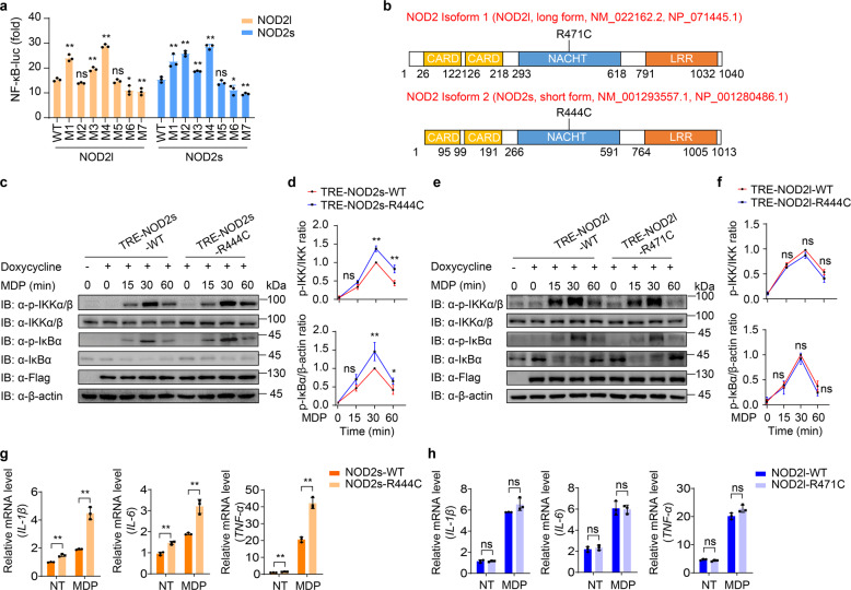
Fig. 4. NOD2s-R444C variant specifically promotes NF-κB activation and inflammation.
a Luciferase activity in 293T cells transfected with the NF-κB luciferase reporter, pRL-TK, together with a vector encoding wild-type (WT) NOD2l, NOD2s, or indicated NOD2 variants (NOD2l: M1, R391W; M2, R471C; M3, G481D; M4, C495Y; M5, R587C; M6, A755V; M7, R702W. NOD2s: M1, R364W; M2, R444C; M3, G454D; M4, C468Y; M5, R560C; M6, A728V; M7, R675W). b Schematic view of NOD2 (full-length/long isoform, NOD2l; short isoform, NOD2s) and the location of the NOD2l-R471C and NOD2s-R444C variants. c–f Immunoblot analysis of muramyl dipeptide (MDP, 10 μg/ml)-induced phosphorylation of IKK and IκBα in NOD2s-WT/R444C (c–d) or NOD2l-WT/R471C (e–f) THP-1-DM cells. Quantitative comparison of signaling activation between WT and NOD2s-R444C (d) or NOD2l-R471C (f) variant cells by density scanning of the immunoblots in three independent experiments. g, h Real-time PCR for IL-1β, IL-6, and TNFα transcription in MDP (10 μg/ml)-induced NOD2s-WT/R444C (g) or NOD2l-WT/R471C (h) THP-1-DM cells. Cells in (c–h) were differentiated with PMA (100 ng/ml) overnight and subsequently treated with Dox (100 ng/ml) for 10 h. In a, d, f, g, and h, all error bars, mean values ± SEM, P values were determined by unpaired two-tailed Student’s t-test of n = 3 independent biological experiments. *P < 0.05; **P < 0.01; ns not significant. For c and e, similar results are obtained from three independent biological experiments.

