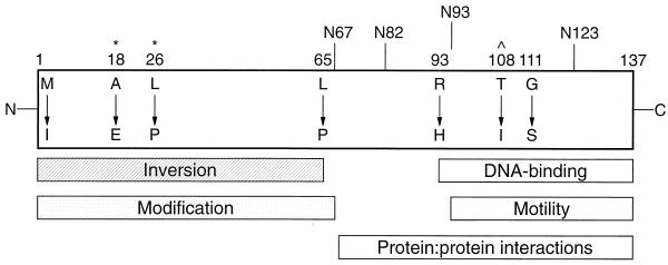
FIG. 4.
Schematic representation of H-NS. Altered residues are indicated by position number and amino acid substitution. Truncations are denoted by vertical lines. Rectangles represent domains involved in the indicated H-NS functions based on this study. The DNA-binding domain is as defined by Ueguchi et al. (42). Caret denotes residue implicated in DNA binding in this study; asterisks represent potential sites of H-NS modification.

