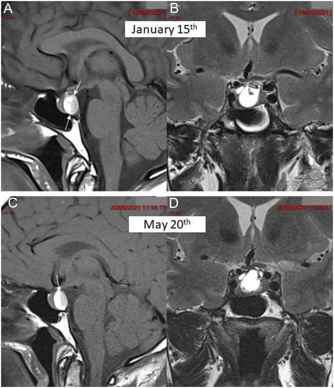
Figure 1.

MRI in patient 1 at the time of diagnosis (upper arrow) and 4 months later (lower arrow). (A) Pre-contrast mid-sagittal T1-weighted view showing enlarged pituitary gland with a posterior cyst disclosing a fluid/fluid level (between arrows) with strongly hyperintense supernatant and a sediment in declivity (patient laying on the back) of moderately lower signal intensity. (B) Coronal T2-weighted view showing the hyperintense liquid content of the cyst with a small hypointense spot at its upper area. (C) Pre-contrast mid-sagittal T1-weighted view in similar slice location as (A) and (D) coronal T2-weighted view in similar slice location as (B) 4 months later failing to demonstrate significant change in the pituitary mass.

 This work is licensed under a