Figure 1.

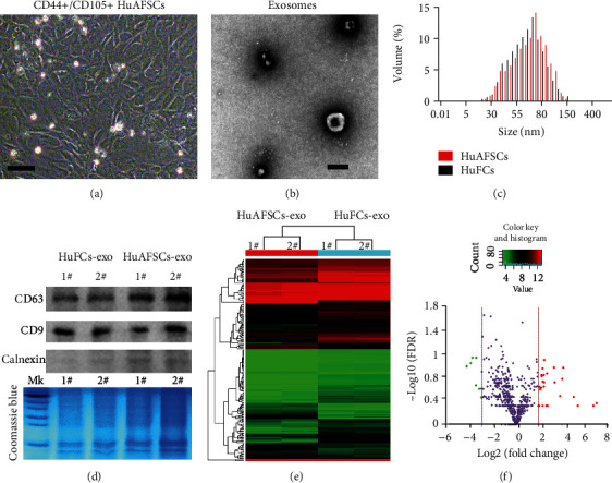
Differences in the microRNA carried by exosomes derived from amniotic fluid stem cells (AFSCs) and exosomes derived from nonstem cells. (a) Morphology of the CD44+/CD105+ subpopulation isolated from human primary amniotic fluid. Original magnification, 200x. (b) Phenotype of HuAFSC-exosomes detected by electron microscopy. Scale bar: 100 nm. (c) The size of HuAFSC-exosomes and HuFCs-exosomes was assessed by nanoFCM. (d) Western blot analysis of exosome marker expression in HuAFSC-exosomes and HuFCs-exosomes. (e) miRNA-Seq results revealed several genes that showed different expression levels between the HuAFSC-exosomes and HuFCs-exosomes. (f) Volcano plot of significant differentially expressed miRNA with a fold-change [log2(HuAFSCs/HuFCs)] > −2 or < ˗3, P < 0.05; green and red colors represent downregulated and upregulated expressions, respectively.
