
Fig. 7.
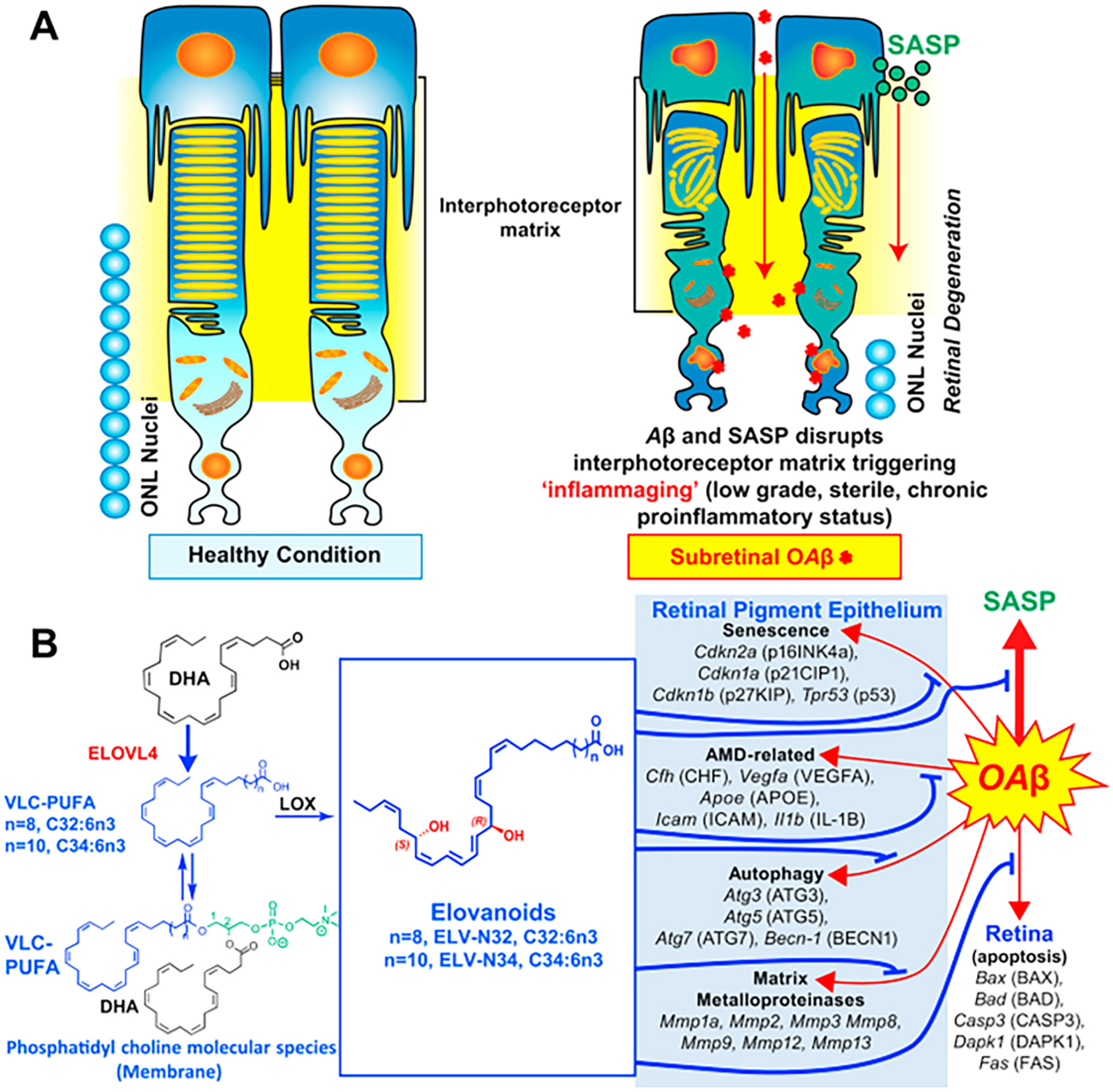
ELVs effect on oligomeric amyloid-β (OAβ)-induced RPE and PRC damage. A: OAβ induces a senescence gene program and disrupts RPE tight junctions. OAβ penetrates the retina, causing PRC cell death in our in vivo WT mice study, as reflected in less outer nuclear layer (ONL) nuclei (Fig. 5 from [198]). OAβ activates the senescence-associated secretome (SASP) that contributes to perturbing the interphotoreceptor matrix (IPM), triggering inflammaging in PRC and also likely in Mueller glia, which limits the IPM. Therefore, senescence paracrine expression takes place. ELVs restore RPE morphology and PRC integrity. B: OAβ induces expression of senescence, autophagy, matrix metalloproteinases, and age-related macular degeneration (AMD)-related genes in the RPE and apoptosis genes in retina in addition to p16INK4a protein abundance. ELVs downregulated the OAβ-gene inductions and p16INK4a protein abundance. Pathways for the ELV synthesis are outlined. ELV, elovanoid; PRC, photoreceptor cell; RPE, retinal pigment epithelium. Reproduced, with permission, from the Proceedings of the National Academy of Sciences of the United States [198].