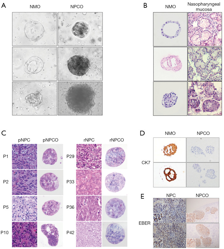
Figure 1.
Morphological and histopathological profiles of NMOs and NPCOs. (A) The morphology of NMOs and NPCOs under light microscopy; (B) H&E staining results for NMOs and their corresponding tissues; (C) H&E staining results for NPCOs and their corresponding NPC tissues; (D) the IHC results for CK7 in NMOs and NPCOs; (E) EBER ISH results for NPCOs and their corresponding NPC tissues. 40× magnification used for organoid models and 20× for tissues. NMO, nasal mucosa organoid; NPCO, nasopharyngeal carcinoma organoid; H&E, hematoxylin and eosin; pNPC, primary NPC; pNPCO, primary NPCO; rNPC, recurrent NPC; rNPCO, recurrent NPCO; IHC, immunohistochemistry; EBER, Epstein-Barr virus (EBV)-encoded small RNAs; ISH, in situ hybridization.

