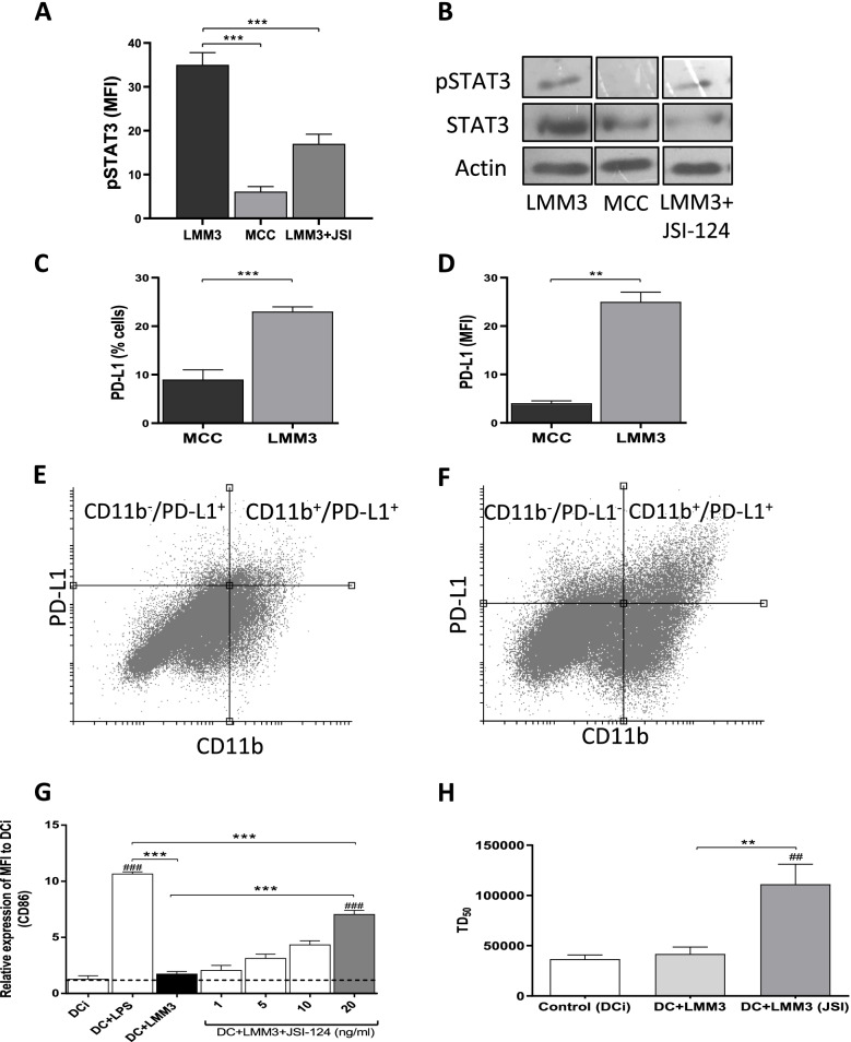
Fig. 2.
Expression of activated STAT3 (pSTAT3) and PD-L1 in MC-C and LMM3 tumors. pSTAT3 was evaluated by flow cytometry (A) and Western Blotting (B) in LMM3, MC-C, and LMM3 tumor cells that have been treated with 20 ng/ml of JSI-124 for 24 h. Controls with actin and total STAT3 were added. Results are representative of three similar experiments. (C, D) Percentage of cells PD-L1+ (C) and mean fluorescence intensity (MFI) (D) in MC-C and LMM3 tumors. Each value represents the mean ± SEM of three assays. (E, F) Representative dot plots of expression of PD-L1 in MC-C (E) and LMM3 (F) tumor and tumor-infiltrating cells. (G) Acquired capacity of LMM3 lysate to promote the maturation of dendritic cells (DC) by pre-treatment with JSI-124. Expression of cell-surface receptor CD86 was evaluated by flow cytometry. DC were incubated with LMM3 lysate or with lysate from LMM3 cells that had been pre-treated in vitro for 24 h with different concentrations (1, 5, 10, and 20 ng/ml) of JSI-124. DC incubated with LPS served as a positive control. Negative controls were immature DC (DCi). Data represent the mean ± SEM of three independent experiments. (H) Vaccinating capacity against LMM3 of DC stimulated with a lysate from LMM3 cells that had been treated in vitro with 20 ng/ml of JSI-124. Two doses of DC of the different groups were inoculated in the footpad of mice 14 and 7 days before the s.c. challenge with different doses of LMM3 tumor cells. Vaccinating capacity was measured as an increase of tumor dose 50 (DT50) of LMM3 tumor in treated mice compared to control. Data represent the mean ± SEM of two independent experiments. In each experiment, 20–25 mice per group were utilized. Statistical comparison between experimental groups and DCi: ## p < 0.01; ### p < 0.001. Statistical comparison among experimental groups: ** p < 0.01; *** p < 0.001

