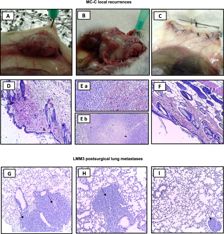
Fig. 6.
Representative macroscopic and histopathological images of tumor recurrences. (A—F) MC-C tumor local recurrences images (H&E staining, 100X) at day 15 post-surgical removal, corresponding to untreated control (A, D), anti-CTLA-4 + anti-PD-L1 (B, E) and anti-CTLA-4 + anti-PD-L1 + m-Tyr + anti-p38 (C, F). Noted the medium-size tumor of untreated mouse (A), the large size tumor of a mouse treated with anti-CTLA-4 + anti-PD-L1 (B), and an imperceptible tumor mass in the suture line from a mouse treated with anti-CTLA-4 + anti-PD-L1 + m-Tyr + anti-p38 (C). (G—I) LMM3 post-surgical lung metastases images (H&E staining, 100X) at day 15 after surgery, corresponding to control (G), anti-CTLA-4 + anti-PD-L1 (H) and anti-CTLA-4 + anti-PD-L1 + m-Tyr + anti-p38 (I) treated mice. Arrows point sites of tumor cells in the same field

