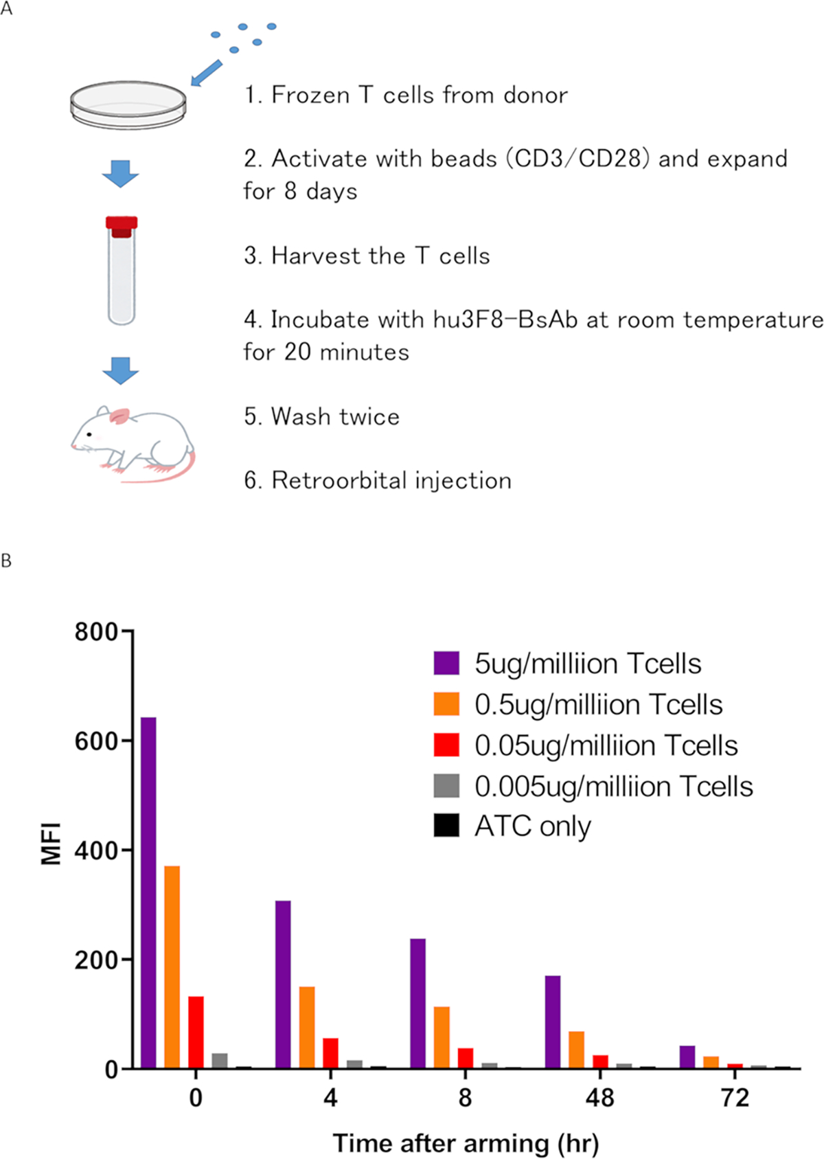
Figure 1. Characteristics of bead activated T cells (ATCs) armed with hu3F8 BsAbs.

(A) The process of expanding T cells ex vivo and arming them with hu3F8 BsAb. (B) Surface hu3F8-BsAb on ATC was assayed using anti-hu3F8 anti-idiotypic antibody A1G4 (labeled with Alexa Fluor 647). Armed ATC was cultured in the absence of hu3F8 BsAb for 4, 8, 24, 48 and 72 hours, washed and stained with A1G4. Surface BsAb was detectable at least through 24 hours after arming. By 72 hours, BsAb has substantially decreased.
MFI, mean fluorescence intensity
