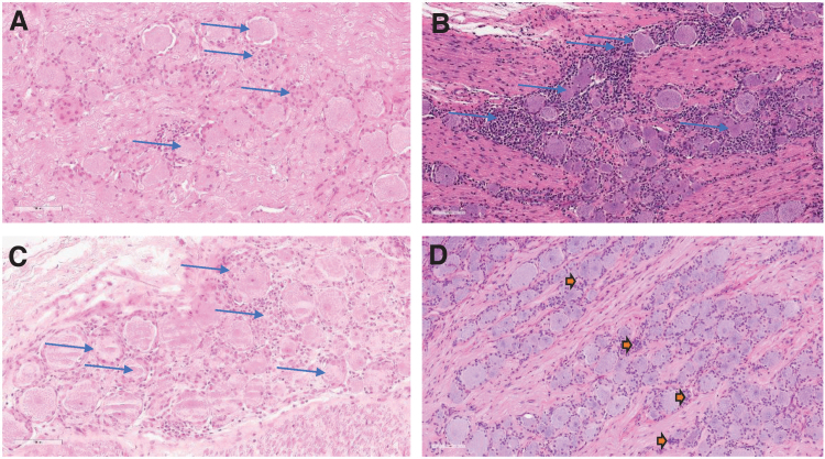
Figure 2.
Microscopic DRG findings at 2, 6, 13, and 52 weeks of observation postdose. H&E-stained, formalin-fixed, paraffin-embedded cervical dorsal root (spinal) ganglia from NHPs dosed 3 × 10¹³ vg/animal by intrathecal lumbar puncture in a dose volume of 0.80 mL. (A) Slight neuron degeneration and mononuclear cell inflammation with minimal increased satellite glial cells at 2 weeks postdose. (B) Moderate neuron degeneration and mononuclear cell inflammation at 6 weeks postdose. (C) Slight neuron degeneration and moderate mononuclear cell inflammation at 13 weeks postdose. (D) Aggregates of satellite glial cells in the cell body rich area of the ganglia at 52 weeks postdose, representing areas of scar or tissue remodeling following neuronal loss because of degeneration. Magnification, 200 × . DRG, dorsal root ganglion; H&E, hematoxylin and eosin.

