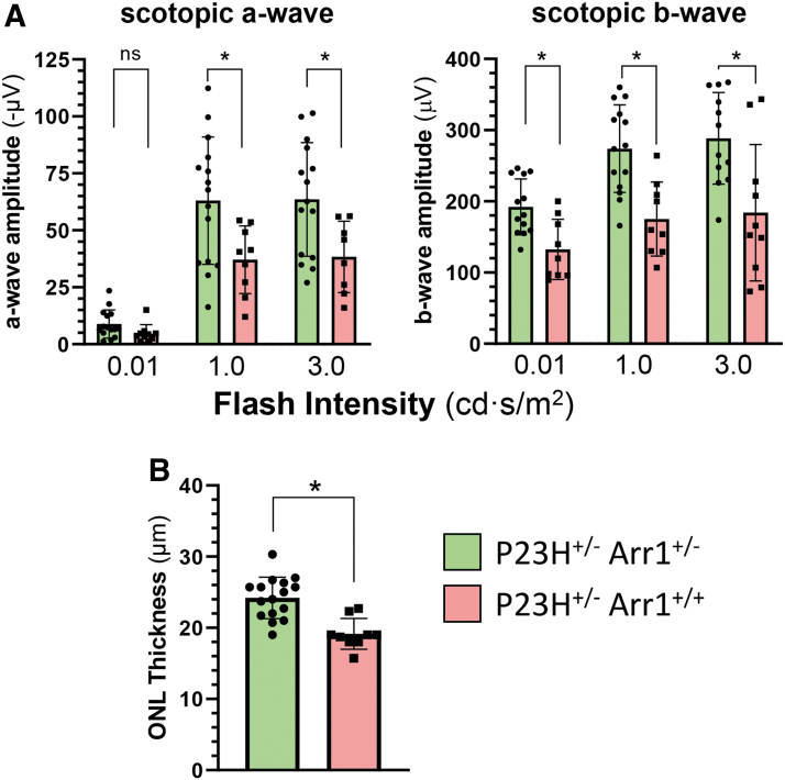
Figure 6.
Photoreceptors are preserved in P23H+/− mice with reduced levels of Arr1 expression. (A) Summary quantitation of scotopic a-wave and b-wave ERG response amplitudes at three flash intensities of a cohort of P23H+/− mice (red bars, n = 9; PN85 days) compared with P23H+/− mice in the background of Arr1+/− (green bars, n = 15; PN100 days). Bars represent mean ± SD, with statistical significance of parametric, unpaired t-test comparisons indicated with an asterisk (ρ < 0.05); ns, not significant. (B) ONL thickness was measured in P23H+/− mice (red bars, n = 9) from OCT images collected at PN92 days and in P23H+/− Arr1+/− mice at PN107 days (green bars, n = 16); ONL measurements are shown at 1,200 μm from the optic nerve head in the temporal retina. Statistical comparison at each time point showed significantly thicker ONL in eyes of P23H+/− mice in the Arr1+/− background compared with eyes from P23H+/− mice in the Arr1+/+ background (bars represent mean ± SD, with statistical significance of parametric, unpaired t-test comparisons indicated with an asterisk (ρ < 0.05). Color images are available online.

