
FIGURE 1.
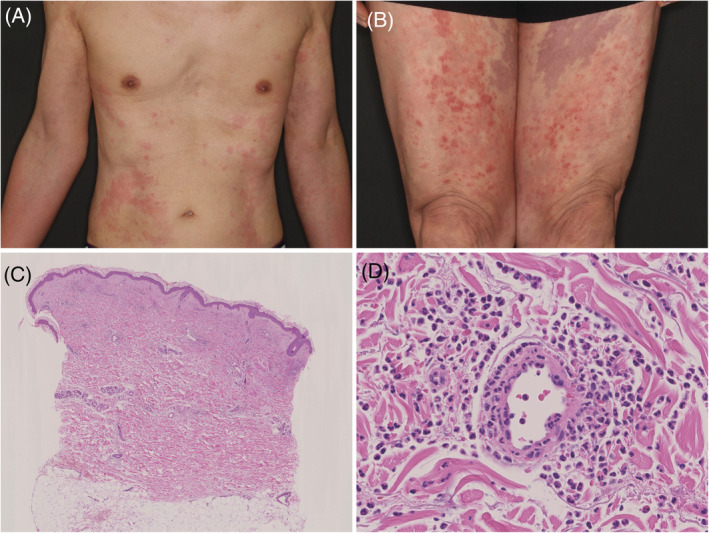
(A) Multiple urticaria‐like skin‐rash lesions on the trunk. (B) Lower extremities covered with sporadic wheals, purpura, and pigmentation lesions. (C) Cellular infiltration was observed perivascular and throughout the dermis (hematoxylin and eosin stain ×100). (D) High‐power view showing perivascular neutrophil infiltration and nuclear debris. Destruction of the vessel wall and erythrocyte leakage are evident (hematoxylin and eosin stain ×400)