
Figure 1.
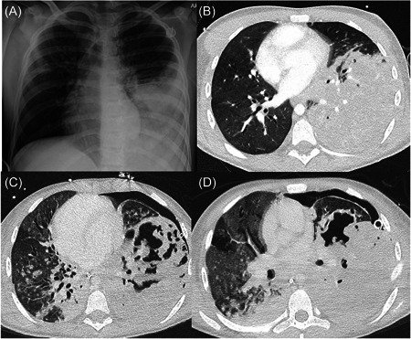
(A) Chest x‐ray on Day 1 of admission showing left lower lobe consolidation. A totally implantable venous access device is also visible. (B) High‐resolution computed tomography (CT) chest on Day 4 of admission showing dense consolidation of the left lower lobe with minimal aerated parenchyma. Right‐sided bronchiectasis also present. (C and D) Repeat high‐resolution CT chest scans undertaken on days 13 and 19 showing left lower lobe necrosis and cavitation. A left‐sided pneumothorax is also seen (different size drains are seen on the two scans).