Figure 1.

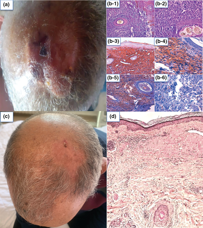
(a) A photo was taken by the patient showing a multinodular tumour of the scalp. (b) Initial histological images: (b‐1) Cutaneous tissue in which the dermis is the site of a lymphomatous proliferation showing a diffuse growth pattern, extending into the hypodermis (HE ×10). (b‐2) Mixture of main centrocytes (large cleaved cells) and centroblasts, provided with irregular nuclei, with one or more prominent nucleoli, arranged within a fibrous stroma (HE ×40). Immunochemical images: (b‐3) Immunochemical labelling CD 20+. (b‐4) Immunochemical labelling Bcl6+. (b‐5) Immunochemical labelling CD10+. (b‐6) Immunochemical labelling Bcl2−. (c) Clinical image showing the disappearance of the scalp tumour after COVID‐19 vaccine, with a residual non‐infiltrated erythema. (d) Histological image after tumour regression showing fibrous scarring.
