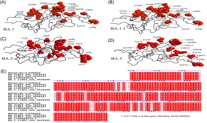
Figure 2.

Ribbon diagram of the RBD with residue mutated relative to the wild‐type (WT). A comparison of (A) Omicron—BA.1, Omicron sub‐variants (B) BA.1.1, (C) BA.2, and (D) BA.3 mutation in receptor‐binding domain (RBD). The multiple alignment (E) of RBD shows receptor‐binding motif (RBM) (residues 437–508) of Omicron variants with WT.
