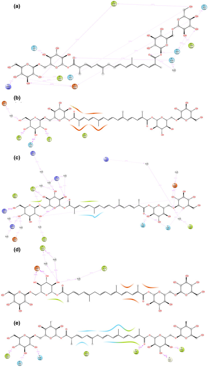
FIGURE 2.

The protein–ligand contacts observed after performing 100 ns MD simulation for all the macromolecular complexes of crocin with (a) Mpro, (b) Helicase, (c) Replication complex, (d) ADRP, and (e) B.1.351 variant of S protein. The macromolecular residues demonstrated in green color have hydrophobic interaction, whereas sky blue‐colored residues have polar interactions with the complexed ligand molecule. The orange‐colored residues are negatively charged, whereas the dark blue residues are positively charged
