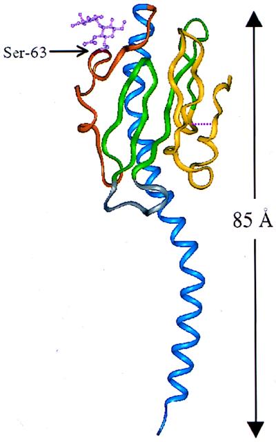
FIG. 1.
Ribbon representation of pilin from N. gonorrhoeae. Colored regions indicate the secondary structural elements referred to in the text: blue, N-terminal α1-helix (residues 2 to 54); orange, extended disaccharide-bound sugar loop (residues 55 to 77); green, β-hairpins (residues 78 to 93 and 103 to 122); gray, β2-β3 loop (residues 94 to 102); yellow, disulfide bond-containing C-terminal region (residues 121 to 158). Also shown are the disulfide bridge (cysteine residues 121 and 151), signified by a broken line, and Ser-63 with covalently linked disaccharide.

