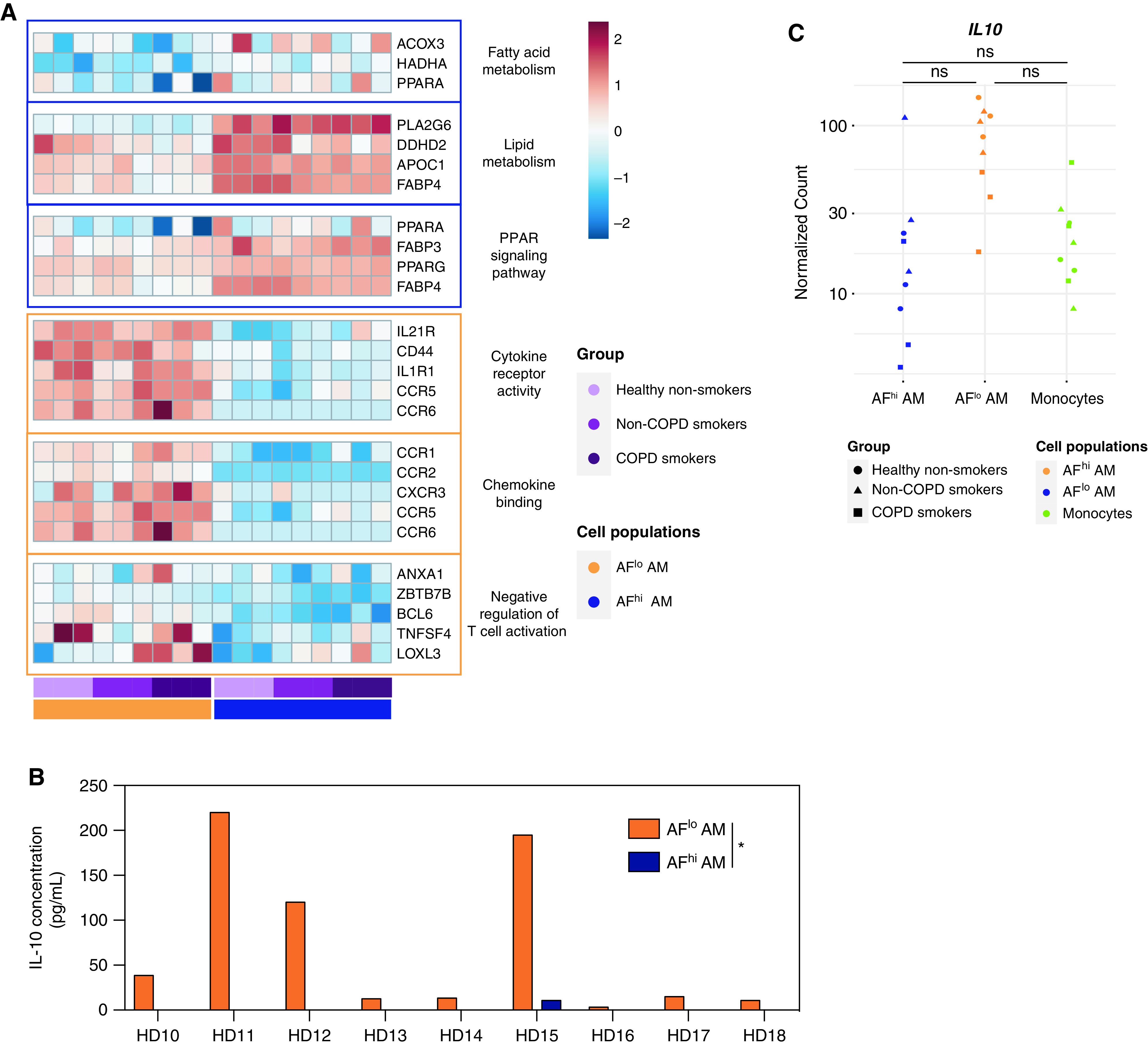
Figure 4.

Monocyte-derived AFlo AMs are functionally distinct from classical AFhi AMs. (A) Heatmaps showing expression of the indicated genes involved in the biological responses that were found to be enriched in AFhi and AFlo AMs analyzed by gene set enrichment analysis (also see Table E3). (B) IL-10 concentrations measured by ELISA in culture supernatant of FACS-sorted AFlo and AFhi AMs from nine patients (HD1 to HD9) (Table E1). (C) Individual expression, shown as normalized counts, of IL-10 gene within the indicated cell populations. Data show individual mean values from technical duplicates. P values were calculated using a two-way ANOVA. *P < 0.05.
