Fig. 2.

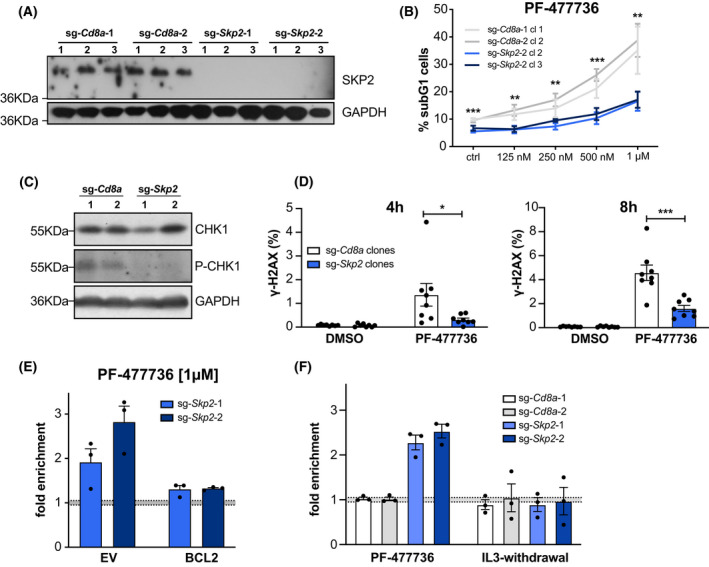
The F‐box protein SKP2 defines cellular sensitivity to CHK1 inhibition. (A) Western blot analysis confirming loss of protein in Baf3 SKP2 KO clones. A single experiment was performed (B) Two randomly selected clones of each genotype were cultured for 48 h in different doses of CHK1‐inhibitor PF‐477736. Cell death was assessed using PI‐staining and flow cytometry. Data points indicate means ± SEM of three independent experiments. (C) Western blot assessing CHK1 and pCHK1Ser345 in SKP2 KO and sg‐Cd8a control clones. (D) Baf3 SKP2 KO‐ and sg‐Cd8a control clones were treated for 4 or 8 h with 1 μm PF‐477736. DNA damage was assessed by γ‐H2A.XSer139 and DAPI staining using flow cytometry. Bars indicate the mean percentage (±SD) of four independent experiments in two clones analyzed per genotype. (E) Baf3 cells expressing pMIP‐BCL2 or an empty vector (EV) control were transduced with two independent sg‐Skp2/dsRed or sg‐Cd8a/dsRed constructs. Three days post transduction the respective bulks were treated with the CHK1 inhibitor PF‐477736 [1 μm] or DMSO for 48 h. The percentage of dsRed+ cells was assessed using flow cytometry, whereby the treatments were normalized to the DMSO‐treated controls. Bars indicate the mean fold enrichment (±SEM) in three independent experiments. (F) Baf3 cells were transduced with two independent sg‐Skp2/dsRed or sg‐Cd8a/dsRed constructs. Three days post transduction the respective bulk cultures were split and treated either with the CHK1 inhibitor PF‐477736 [1 μm] or DMSO for 48 h (left panel); or depleted for IL‐3 or left untreated for 48 h (right panel). The percentage of dsRed+ cells was assessed using flow cytometry, whereby the PF‐477736‐treated cells were normalized to the DMSO‐treated controls, and the IL‐3‐depleted cells were normalized to the untreated controls. Bars indicate the mean fold enrichment (±SEM) noted in three independent experiments. *P < 0.05; **P < 0.01; ***P < 0.001; unpaired t‐test.
