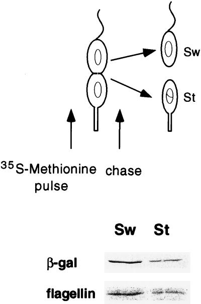
FIG. 4.
Cell type-specific expression of hfaA. The transcriptional fusion containing the hfaA promoter region (pRJ38) is shown in Fig. 3A. Cells containing pRJ38 were synchronized, and the swarmer cells were allowed to proceed to the late-predivisional stage (135 min). Proteins were labeled with [35S]methionine for 10 min and then chased with unlabeled methionine as shown. After division, the stalked (St) and swarmer (Sw) cells were separated by density gradient centrifugation and then processed as shown in Fig. 3. The autoradiograph of immunoprecipitated β-galactosidase and flagellin proteins is shown. This experiment was repeated twice with reproducible results.

