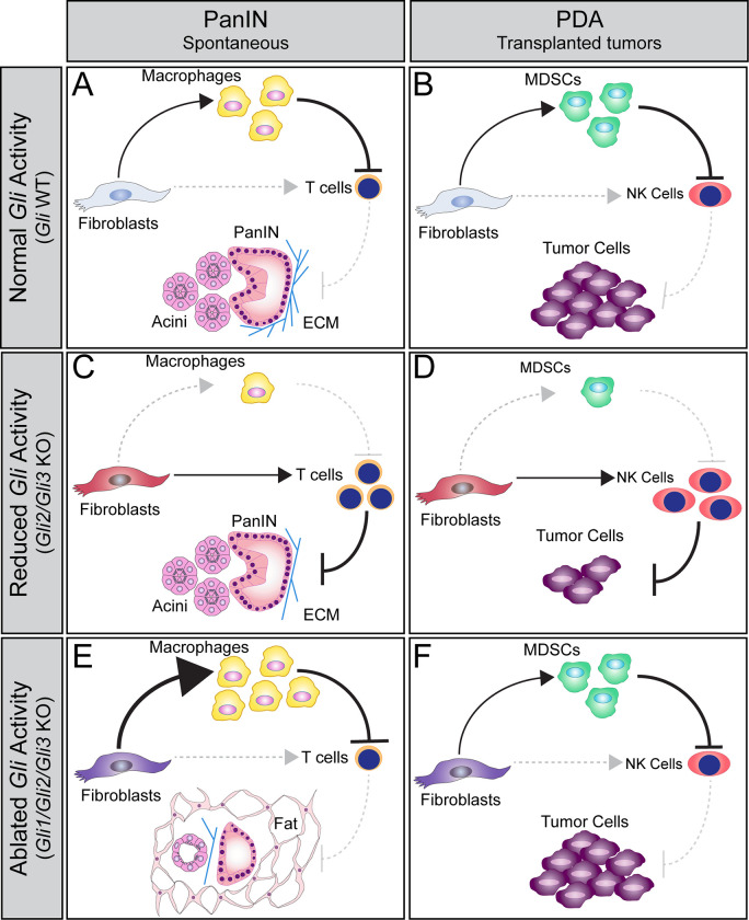
Fig 7. Model of GLI function during PDA progression.
(A-B) Gli-expressing fibroblasts directly promote the recruitment of macrophages and MDSCs (A,B, top) at PanIN and PDA stages, respectively. These myeloid cells suppress T cells and NK cells (A,B, right), facilitating disease progression. (C-D) Gli2 and Gli3 deletion in fibroblasts directly reduces the recruitment of myeloid cells, and directly promotes T cell and NK cell infiltration (C,D, right). Loss of Gli2 and Gli3 also decreases collagen deposition and slows tumor growth (C,D, bottom). However, when all three Glis are deleted (E, F), fibroblasts have an enhanced ability to recruit macrophages (E, top) and a sustained ability to recruit MDSCs (F, top), leading to T cell and NK cell exclusion (E,F, right). Thus, Gli1/Gli2/Gli3 KO fibroblasts support tumor growth (F, bottom). Interestingly, loss of Gli1/Gli2/Gli3 also leads to the loss of pancreas tissue and an accumulation of fat at PanIN stages (E, bottom), indicating that a baseline level of GLI activity is necessary to maintain pancreas integrity.

