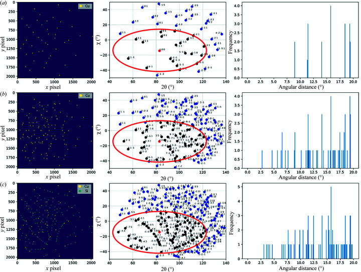
Figure 3.
Simulated Laue image, ground truth indexing and angular distribution around a single peak for (a) a Cu single crystal, (b) Cu polycrystals (three crystals), and (c) Cu and Si single crystals. The leftmost subplots present the Laue spots in pixel coordinates, the middle plots present the Laue spots in scattering angle coordinates (the ellipse is a guide for eye to represent the spots within 20° of angular distance from the red spot), and the rightmost plots are the histograms of radial angular distributions (with fixed bins of 0.1°) of neighbouring spots around a single Laue spot.

