Figure 1.

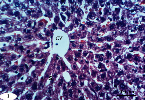
Photomicrograph of liver section in an untreated rat indicating normal liver architecture composed of a central vein (CV) with thin walls and normal hepatocytes with narrow intercellular sinusoids (S) (H&E; ×400).

Photomicrograph of liver section in an untreated rat indicating normal liver architecture composed of a central vein (CV) with thin walls and normal hepatocytes with narrow intercellular sinusoids (S) (H&E; ×400).