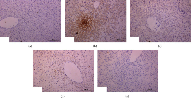
Figure 7.

Photomicrographs of immunohistochemical sections of liver for detection of TNF-α showing weak expression of in normal rats (a), strong expression (immunopositivity indicated by brownish color) in DXR-injected rats (b), weak expression in DXR-injected rats treated with rutin (c) and quercetin (d), and negative expression in DXR-injected rats treated with mixture of rutin and quercetin (e) (×100).
