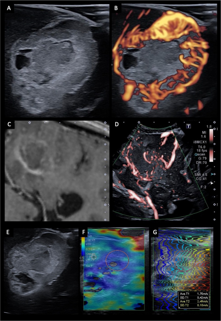
Fig. 5.
Advanced ultrasound imaging techniques. Examples of US doppler using superb microvascular imaging (SMI) (A-D) and elastography (E–G). B-mode (A) and doppler (B) imaging of a glioblastoma shows a relatively well-circumscribed, hyperechoic tumor with small hypoechoic areas in keeping with cystic necrosis (A). The tumor exhibits marked peripheral hypervascularity in keeping with neoangiogenesis (B). Post-contrast T1-weighted MRI (C) and doppler imaging (D) of a glioblastoma demonstrates the ability of US doppler imaging to resolve and identify small pial and parenchymal vessels (red) that are not appreciable on MRI. B-mode US imaging (E) and shear wave elastography (F, G) of a glioblastoma. The tumor demonstrates reduced stiffness compared to the surrounding brain, which is reflected by a heat map with blue reflecting low stiffness and red high stiffness

