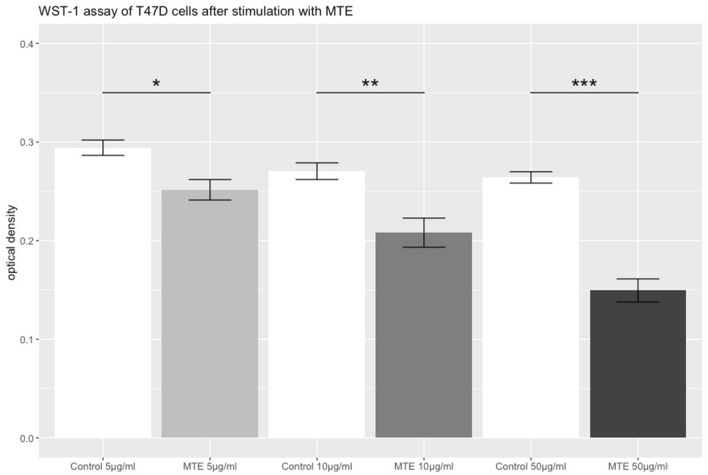
Fig. 2.
WST-1 assay of T47D cells stimulated with MTE. The grey bars represent the optical density of T47D cells after the incubation with different concentrations of MTE (5, 10 and 50 µg/ml) for 72 h. The white bars represent the control group. The top of each bar represents the mean ± standard error (SE). MTE induced a significant reduction of cell proliferation at every concentration. Significant results are linked and marked with asterisks (p < 0.05*, p < 0.01**, p < 0.001***)

